
Conditions We Treat
PEMF for Tendon &
Ligament Injuries
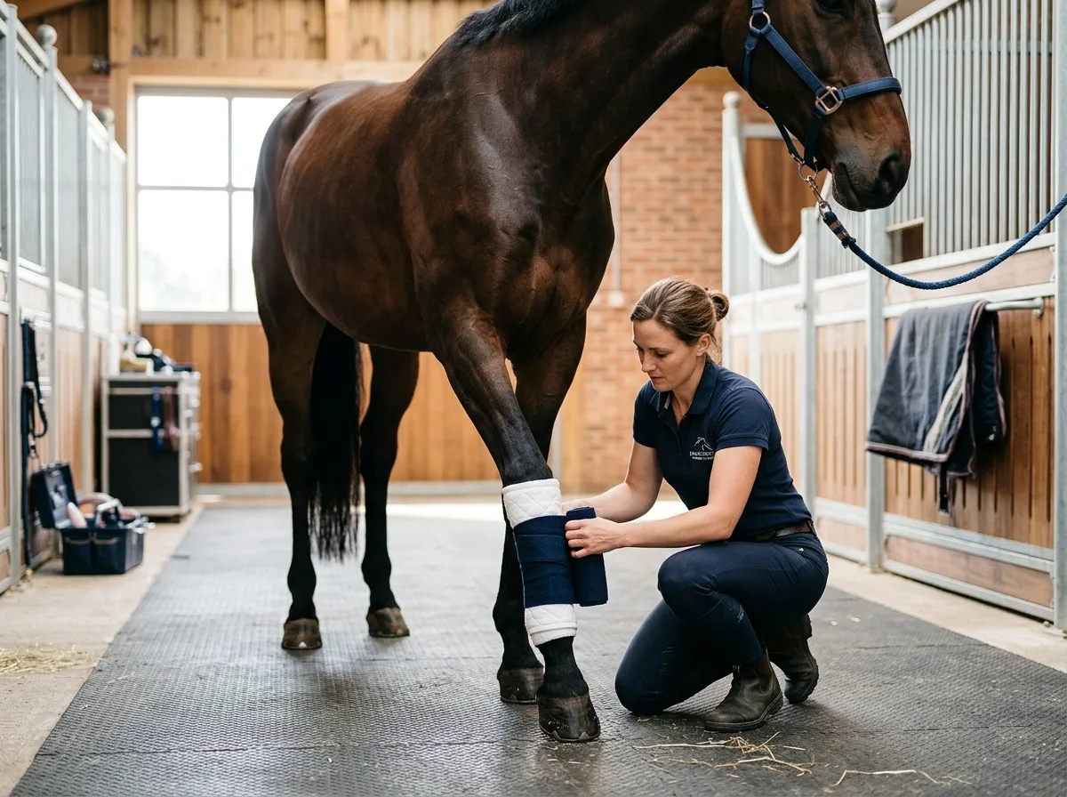
Tendon and ligament injuries are among the most frustrating challenges horse owners face. These soft tissue structures heal slowly, are prone to re-injury, and require extended periods of controlled rest. PEMF therapy has become an increasingly valued part of equine rehabilitation programs because it directly supports the biological processes that drive tissue repair, helping horses heal more completely and return to work with stronger, better-organized tissue.
Common Equine Soft Tissue Injuries
The equine lower limb is a marvel of engineering, with tendons and ligaments doing the heavy lifting of supporting body weight, absorbing concussion, and storing elastic energy for locomotion. But this design also means these structures are under enormous stress, making injury a persistent risk — especially in athletic horses.
- Superficial digital flexor tendon (SDFT) injuries: Often called a "bowed tendon," this is the most common tendon injury in sport horses. The SDFT runs down the back of the cannon bone and is responsible for supporting the fetlock during weight-bearing. Injury results in swelling, heat, pain, and the characteristic outward bowing of the tendon sheath. Racing Thoroughbreds, eventers, and show jumpers are particularly susceptible.
- Deep digital flexor tendon (DDFT) injuries: The DDFT runs alongside the SDFT but inserts on the bottom of the coffin bone inside the hoof. Injuries to this tendon can be harder to diagnose because the swelling may be less visible, but they cause significant lameness and can involve the tendon where it passes through the navicular region.
- Suspensory ligament injuries: The suspensory ligament supports the fetlock joint from behind and is critical for preventing hyperextension during movement. Injuries can occur at the origin (top), body (middle), or branches (bottom) of the ligament, with each location presenting different challenges for healing and prognosis.
- Check ligament injuries: The inferior and superior check ligaments work in conjunction with the flexor tendons. Injuries to these structures are less common but can be debilitating and often occur alongside other soft tissue damage.
- Collateral ligament injuries: The collateral ligaments stabilize joints laterally, and injuries — particularly in the coffin joint and fetlock — can cause persistent lameness that is sometimes difficult to localize without advanced imaging.
Why Tendon and Ligament Injuries Heal Slowly
Tendons and ligaments have inherently poor blood supply compared to muscle tissue. This limited vascularity means fewer nutrients and oxygen reach the damaged area, and the cellular processes of repair happen at a slower pace. Additionally, the body's initial repair response produces scar tissue (type III collagen) rather than the stronger, more organized type I collagen that makes up healthy tendon. This scar tissue is weaker, less elastic, and more prone to re-injury — which is why recurrence rates for tendon injuries remain high even with careful rehabilitation.
Healing timelines for significant tendon and ligament injuries typically range from six months to over a year, with the first three months being the most critical for tissue repair and the remaining time devoted to remodeling and strengthening the new tissue. Rushing the process is one of the primary reasons for re-injury.
How PEMF Accelerates Soft Tissue Healing
PEMF therapy addresses the fundamental limitations of tendon and ligament healing by enhancing the biological environment in which repair occurs:
- Enhanced blood flow: PEMF therapy promotes vasodilation and angiogenesis (the formation of new blood vessels) in the treated area. For tendons and ligaments with naturally poor blood supply, this increased circulation is transformative — bringing more oxygen, growth factors, and nutrients to the repair site while removing waste products more efficiently.
- Reduced edema and swelling: Swelling compresses the tissue, restricts blood flow, and impedes healing. PEMF helps move excess fluid out of the injured area, reducing pressure on the tissue and creating a better environment for repair.
- Improved collagen organization: Research suggests that PEMF therapy can influence the alignment and organization of collagen fibers during the repair process. Better-organized collagen produces stronger tissue with more elasticity — closer to the original tendon or ligament structure rather than disorganized scar tissue.
- Increased cellular energy: By stimulating ATP production in fibroblasts (the cells responsible for building collagen), PEMF accelerates the rate at which new tissue is produced. More active fibroblasts mean faster, more robust repair.
- Anti-inflammatory effects: Chronic inflammation around an injured tendon can actually slow healing by maintaining tissue in a destructive rather than reparative state. PEMF helps shift the balance toward productive healing by moderating inflammatory mediators.
Working Alongside Veterinary Rehabilitation
Tendon and ligament injuries require veterinary management, and PEMF therapy is most effective when integrated into a comprehensive rehabilitation program designed by your veterinarian. A typical program includes initial assessment with diagnostic ultrasound to determine the extent and location of damage, a controlled exercise protocol that gradually increases load on the healing tissue, regular ultrasound re-checks to monitor tissue repair, and complementary therapies like PEMF that support the healing process at each stage.
PEMF therapy does not replace stall rest when your veterinarian has prescribed it. The controlled restriction of movement during the acute healing phase is essential to prevent further damage. What PEMF does is make that healing time more productive by enhancing the quality and speed of tissue repair within the safety of a controlled rehabilitation program.
Treatment Protocol for Soft Tissue Injuries
For acute injuries, we typically begin PEMF therapy within the first week after veterinary assessment, once the initial acute inflammation has been managed. Sessions during the early healing phase focus on reducing swelling and pain while supporting the initial cellular repair response. We recommend sessions every three to five days during the first month.
As healing progresses into the proliferative phase (typically weeks four through twelve), we continue with weekly sessions to support collagen production and organization. During the remodeling phase (three months onward), sessions can be spaced to every two to three weeks, though many owners choose to maintain regular sessions through the entire return-to-work protocol.
Each session lasts approximately 30 to 40 minutes. We focus the PEMF applicator directly on the injured structure while also treating the surrounding musculature and compensatory areas — because a horse protecting an injured leg inevitably develops tension elsewhere. The whole body benefits from treatment even when one area is the primary focus.
Expected Outcomes
Owners who incorporate PEMF therapy into their horse's tendon or ligament rehabilitation commonly report reduced swelling sooner than expected, improved comfort and attitude during the stall rest period, positive ultrasonographic findings at re-check appointments showing good fiber alignment, and smoother transitions as the horse returns to increasing levels of work. While outcomes vary with the severity and location of the injury, PEMF therapy consistently contributes to a better healing environment and a more complete recovery.
Related Conditions
Quick Facts
- Condition
- Tendon & ligament injuries (SDFT, DDFT, suspensory)
- Primary Benefit
- Accelerated tissue repair with better collagen organization
- Session Length
- 30 – 40 minutes
- Acute Phase
- Every 3 – 5 days for the first month
- Rehab Phase
- Weekly through months 2 – 3
- Important Note
- Works alongside vet-prescribed rest — never a substitute
Dealing with a soft tissue injury?
PEMF can accelerate your horse's recovery. We bring the treatment to your barn.
Book a Session